|
Ear
Reconstruction
Mr Davis has studied with internationally recognized leaders in
ear reconstruction including Dr Francois Firmin (France), Dr
David Gault (UK), Dr
S O Wickstrom (Sweden), Dr
Burt Brent (USA), Dr
David Fisher and Dr
John Vandervord (Australia). His current technique is a mosaic
of each of these experts techniques and he has performed ear reconstructions
in Sweden, Australia, Sri Lanka and Malaysia as well as New Zealand.
|
BEFORE |
AFTER |
|
 |
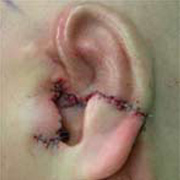 |
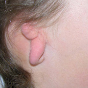
Lop ear
Lop ear after the first stage of reconstruction |
 |
 |
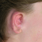
Meurmann Grade III microtia
Meurmann Grade III microtia after 1st stage reconstruction
|
 |
 |
Meurmann Grade III microtia
Meurmann Grade III microtia prior to elevation of framework |
 |
 |
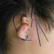
Meurmann Grade III microtia
Meurmann Grade III microtia prior to framework elevation.
Note position of BAHA well away from reconstructed ear. |
 |
 |
Meurmann Grade III microtia
Meurmann Grade III microtia after 1st stage reconstruction |
 |
 |
Meurmann Grade II microtia
Meurmann Grade II microtia soon after framework elevation.
Swelling obliterates the definition of the framework for many
weeks after surgery during the second stage. |
 |
 |
Ear reconstruction
Ear reconstruction with tissue expanded flap and carved costal
cartilage after severe otoplasty complication in another hospital. |
 |
 |
Ear reconstruction
Ear reconstruction with carved costal cartilage after dog
bite |
|
|
Click here for a PDF
about the technique used in first stage microtia correction >>
What are the usual causes of an ear anomaly?
Ear anomalies result from either a deformation or a developmental
malformation. Deformational ear anomalies are best treated
non-surgically with a splint within the first 3 months of life.
Malformations (including microtia and anotia) and
ear defects following trauma such as a dog bite or a burn are best
treated surgically. Anotia is complete absence of the outer ear
at birth and microtia is the presence of a small ear or ear remnants.
Microtia occurs in about 1 in every 3000 births and it may be isolated
or part of a syndrome such as hemifacial microsomia, Goldenhar syndrome
or Treacher Collins syndrome. There are about 20 new cases of microtia
in New Zealand each year.
What is the purpose of an ear reconstruction?
An ear reconstruction aims to rebuild an external ear that is closer
to normal in appearance. At a quick glance to a casual observer
it should look like an ear and not catch the eye in the way an untreated
ear might. Microtia is often associated with abnormalities of the
middle ear and hearing impairment.
Outer ear reconstruction does not improve hearing.
30-40% of patients with facial deformity experience psychological
distress at some time and this is usually relieved with reconstructive
surgery. Ear reconstruction is also useful for those who wear glasses.
What is involved in an ear reconstruction?
The new ear is made from the patients own tissues and requires at
least 2 operations. In the first operation cartilage is taken from
the rib cage and carved to create a framework that resembles a normal
ear. The other ear is used as a template if it is normal. This cartilage
framework is inserted beneath a pocket of skin. This operation takes
3-4 hours and the child stays in hospital for about 2-3 days. At
this stage, the shape of the new ear can be seen but it will lie
flat against the side of the head.
At the second operation the ear framework is elevated
and the groove behind the ear created. This is normally done at
least 4 months after the first operation to ensure a good blood
supply to the cartilage framework. A flap of tissue from the scalp
covers the back of the cartilage which is in turn covered by a skin
graft. This operation takes about 3 hours and the child stays in
hospital for 2 days.
The new reconstructed ear is quite stiff and does
not bend like a normal ear. However it is warm and will eventually
get some feeling. Possible complications may include the need for
revisions. It is important to realize that the new ear will not
be exactly like a normal ear.
What is the timing of surgery?
By age 4, the ear is nearly 85% of adult size. However the chest
is not sufficiently grown and most ear reconstructions are usually
postponed until about age 10 or older. The advantage of waiting
is to ensure sufficient cartilage for the framework and less growth
disturbance of the chest wall. Earlier surgery is considered when
the child is suffering extreme psychological distress.
What about surgery to improve hearing?
Potential candidates for middle ear surgery are seen conjointly
at the Microtia clinic with Dr Rebecca Garland, a consultant ENT
surgeon who specialises in Paediatric Otology.
Reconstruction of the hearing mechanism and ear
canal is possible in some children who have bilateral microtia.
The new Bone Anchored Hearing aid system may be available for those
children who are not suitable for reconstruction, or if their parents
prefer this. On some occasions children with only one ear affected
may be suitable for a bone anchored hearing aid. Assessment of hearing
in the classroom by education department staff is recommended before
referral for this. Children who have a tiny ear canal (“pin
hole”) should have a CT scan of the ears at age 3 to exclude
the possibility of cholesteatoma, a destructive build up of skin
cells internally.
Are there other options available?
A prosthetic ear may be created using the normal ear as a template.
Two operations will be required. At the first stage any ear remnants
are removed and 3 titanium implants are embedded into the bone on
the side of the head. At the second operation, usually 4 months
later, a metal frame is attached to these implants. The prosthetic
ear is clipped onto this metal frame.
A prosthetic ear will require regular maintenance and needs replacement
every two years. The area around the implants must be cleaned daily
and the prosthetic ear is generally removed at night. This can be
a good option for adults with extensive local scarring such as burns
where the local tissue may not be ideal for a surgical reconstruction.
|
